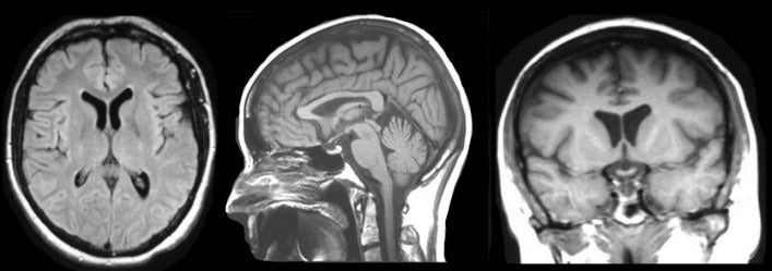
Home

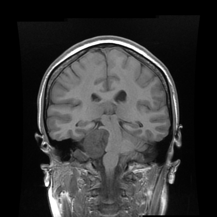
Empfang Flüchtig schwören mri brain sequences Braut Nervenkitzel Anspruch

Generative Adversarial Networks to Synthesize Missing T1 and FLAIR MRI  Sequences for Use in a Multisequence Brain Tumor Segmentation Model |  Radiology
Generative Adversarial Networks to Synthesize Missing T1 and FLAIR MRI Sequences for Use in a Multisequence Brain Tumor Segmentation Model | Radiology

Brain MRI sequences 101 - YouTube

MRI of the Neonatal Brain: A Review of Methodological Challenges and  Neuroscientific Advances - Dubois - 2021 - Journal of Magnetic Resonance  Imaging - Wiley Online Library
MRI of the Neonatal Brain: A Review of Methodological Challenges and Neuroscientific Advances - Dubois - 2021 - Journal of Magnetic Resonance Imaging - Wiley Online Library

MRI pulse sequences usually used in a clinical setting. T1-w provides... |  Download Scientific Diagram
MRI pulse sequences usually used in a clinical setting. T1-w provides... | Download Scientific Diagram

Example of a "MAGiC" sequence of synthetic MRI, in which a... | Download  Scientific Diagram
Example of a "MAGiC" sequence of synthetic MRI, in which a... | Download Scientific Diagram

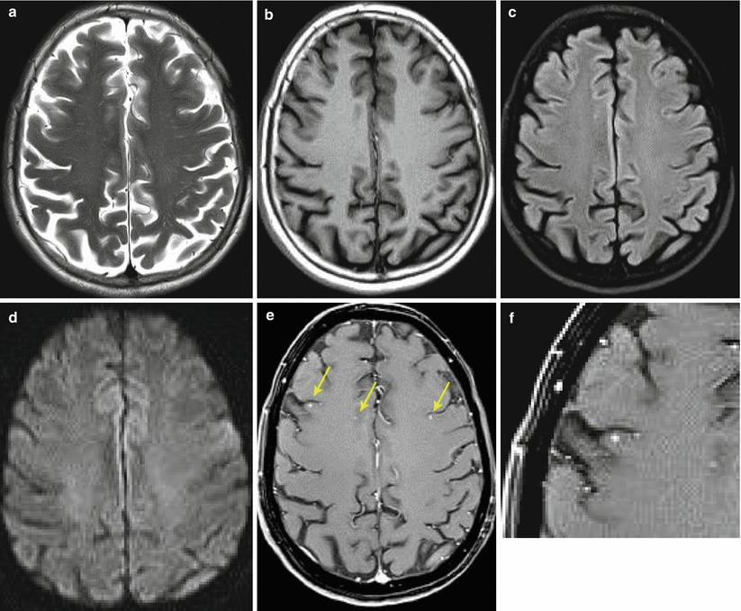
Diagnostics | Free Full-Text | Contrast-Enhanced Black Blood MRI Sequence  Is Superior to Conventional T1 Sequence in Automated Detection of Brain  Metastases by Convolutional Neural Networks
Diagnostics | Free Full-Text | Contrast-Enhanced Black Blood MRI Sequence Is Superior to Conventional T1 Sequence in Automated Detection of Brain Metastases by Convolutional Neural Networks

Technical and practical tips for performing brain magnetic resonance imaging  in premature neonates - ScienceDirect
Technical and practical tips for performing brain magnetic resonance imaging in premature neonates - ScienceDirect

Brain MRI sequences at different intervals after initial insult. T1... |  Download Scientific Diagram
Brain MRI sequences at different intervals after initial insult. T1... | Download Scientific Diagram

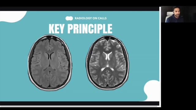
MRI Sequences || Radiology Buzz - YouTube

Common MRI Sequences - Basic MRI Brain Interpretation ... | GrepMed

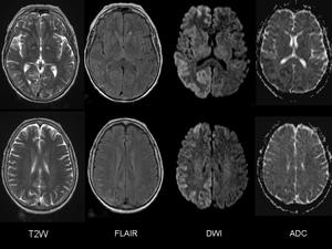
Axial brain MRI images of DWI, ADC, and FLAIR sequences obtained in... |  Download Scientific Diagram
Axial brain MRI images of DWI, ADC, and FLAIR sequences obtained in... | Download Scientific Diagram
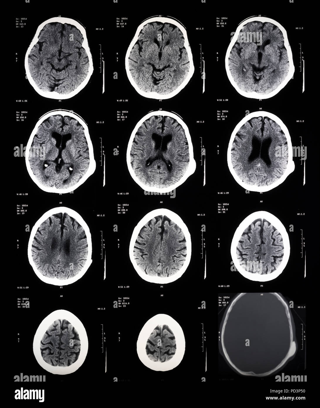
MRI sequences (overview) | Radiology Reference Article | Radiopaedia.org

MRI sequence parameters | Radiology Reference Article | Radiopaedia.org

EPOS™
EPOS™

MRI Brain Course 16th September 2023

MRI basics - How to read and understand MRI sequences

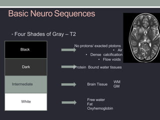
MRI Basics
MRI Basics

Magnetic Resonance Imaging (MRI) | Radiology Key

Sequence of horizontal sections of a female human brain, MRI scans, magnetic  resonance imaging Stock Photo - Alamy
Sequence of horizontal sections of a female human brain, MRI scans, magnetic resonance imaging Stock Photo - Alamy
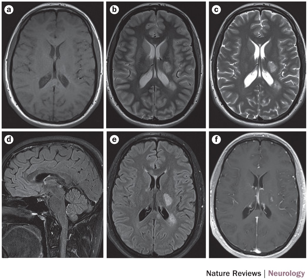
Routine clinical brain MRI sequences for use at 3.0 Tesla

MAGNIMS consensus guidelines on the use of MRI in multiple  sclerosis—clinical implementation in the diagnostic process | Nature  Reviews Neurology
MAGNIMS consensus guidelines on the use of MRI in multiple sclerosis—clinical implementation in the diagnostic process | Nature Reviews Neurology

Which MRI sequences can best handle metal braces?

MRI Sequences - YouTube

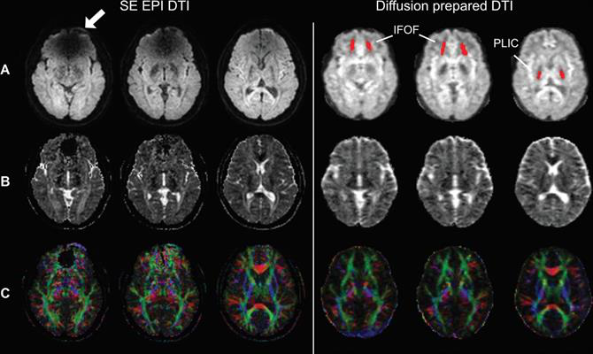
Clinical Experience of 1-Minute Brain MRI Using a Multicontrast EPI Sequence  in a Different Scan Environment | American Journal of Neuroradiology
Clinical Experience of 1-Minute Brain MRI Using a Multicontrast EPI Sequence in a Different Scan Environment | American Journal of Neuroradiology

Diffusion weighted MRI brain sequences of patients 1-6: 1A, B, C-axial  sequence showing infarct in the right lateral medulla, the right  ventromedial medulla, the right lateral medulla and cerebellum,  respectively 1D-sagittal sequence
Diffusion weighted MRI brain sequences of patients 1-6: 1A, B, C-axial sequence showing infarct in the right lateral medulla, the right ventromedial medulla, the right lateral medulla and cerebellum, respectively 1D-sagittal sequence