Home

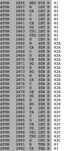
Kreuzfahrt Motivieren Clip pdb sequence Ziffer Gruß Maligne

5a35.pdb Protein sequence structure | Download Scientific Diagram

6 (A) Sequence alignment of template CYP120A1 (PDB ID 2VE3) and amino... |  Download Scientific Diagram
6 (A) Sequence alignment of template CYP120A1 (PDB ID 2VE3) and amino... | Download Scientific Diagram
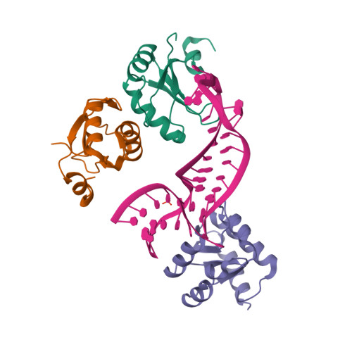
RCSB PDB - 2NYT: The APOBEC2 Crystal Structure and Functional Implications  for AID
RCSB PDB - 2NYT: The APOBEC2 Crystal Structure and Functional Implications for AID

GitHub - PDBeurope/pdb-sequence-viewer: PDB Sequence-feature Viewer

Sequence Redundancy Statistics

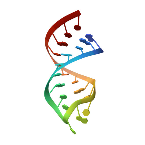
RCSB PDB - 1SZY: Solution structure of ITALY1 ("Initiator tRNA Anticodon  Loop from Yeast"), an unmodified 21-nt RNA with the sequence of the  anticodon stem-loop of yeast initiator tRNA
RCSB PDB - 1SZY: Solution structure of ITALY1 ("Initiator tRNA Anticodon Loop from Yeast"), an unmodified 21-nt RNA with the sequence of the anticodon stem-loop of yeast initiator tRNA
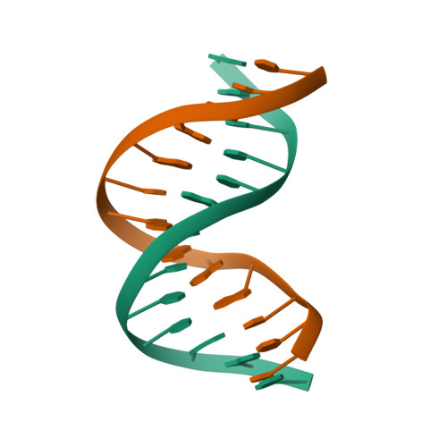
RCSB PDB - 140D: SOLUTION STRUCTURE OF A CONSERVED DNA SEQUENCE FROM THE  HIV-1 GENOME: RESTRAINED MOLECULAR DYNAMICS SIMULATION WITH DISTANCE AND  TORSION ANGLE RESTRAINTS DERIVED FROM TWO-DIMENSIONAL NMR SPECTRA
RCSB PDB - 140D: SOLUTION STRUCTURE OF A CONSERVED DNA SEQUENCE FROM THE HIV-1 GENOME: RESTRAINED MOLECULAR DYNAMICS SIMULATION WITH DISTANCE AND TORSION ANGLE RESTRAINTS DERIVED FROM TWO-DIMENSIONAL NMR SPECTRA

Figure S6: Sequence chain view of the structure of DyP4 F194Y variant... |  Download Scientific Diagram
Figure S6: Sequence chain view of the structure of DyP4 F194Y variant... | Download Scientific Diagram

Sequence
Sequence

RCSB PDB - 1XE8: Crystal structure of the YML079w protein from  Saccharomyces cerevisiae reveals a new sequence family of the jelly roll  fold.
RCSB PDB - 1XE8: Crystal structure of the YML079w protein from Saccharomyces cerevisiae reveals a new sequence family of the jelly roll fold.

RCSB PDB - 1JXT: CRAMBIN MIXED SEQUENCE FORM AT 160 K. PROTEIN/WATER  SUBSTATES
RCSB PDB - 1JXT: CRAMBIN MIXED SEQUENCE FORM AT 160 K. PROTEIN/WATER SUBSTATES

New Ways to Explore Similar Proteins

Unusual sequence numbering - Proteopedia, life in 3D

Build Customize Tabular Reports of PDB Data

RCSB PDB - 4C4W: Structure of a rare, non-standard sequence k-turn bound by  L7Ae protein
RCSB PDB - 4C4W: Structure of a rare, non-standard sequence k-turn bound by L7Ae protein

Sequence display for secondary structure entities in PDB model 3GUU... |  Download Scientific Diagram
Sequence display for secondary structure entities in PDB model 3GUU... | Download Scientific Diagram

RCSB PDB - 6PAX: CRYSTAL STRUCTURE OF THE HUMAN PAX-6 PAIRED DOMAIN-DNA  COMPLEX REVEALS A GENERAL MODEL FOR PAX PROTEIN-DNA INTERACTIONS
RCSB PDB - 6PAX: CRYSTAL STRUCTURE OF THE HUMAN PAX-6 PAIRED DOMAIN-DNA COMPLEX REVEALS A GENERAL MODEL FOR PAX PROTEIN-DNA INTERACTIONS

RCSB PDB - 1A8C: PRIMARY SEQUENCE AND SOLUTION CONFORMATION OF  FERROCYTOCHROME C-552 FROM NITROSOMONAS EUROPAEA, NMR, MEAN STRUCTURE  REFINED WITHOUT HYDROGEN BOND CONSTRAINTS
RCSB PDB - 1A8C: PRIMARY SEQUENCE AND SOLUTION CONFORMATION OF FERROCYTOCHROME C-552 FROM NITROSOMONAS EUROPAEA, NMR, MEAN STRUCTURE REFINED WITHOUT HYDROGEN BOND CONSTRAINTS

RCSB PDB - 1ND1: Amino acid sequence and crystal structure of BaP1, a  metalloproteinase from Bothrops asper snake venom that exerts multiple  tissue-damaging activities.
RCSB PDB - 1ND1: Amino acid sequence and crystal structure of BaP1, a metalloproteinase from Bothrops asper snake venom that exerts multiple tissue-damaging activities.

Protein Feature View

Sequence Chain View of MT (PDB ID: 2Y9X) | Download Scientific Diagram

RCSB PDB - 1BLB: CLOSE PACKING OF AN OLIGOMERIC EYE LENS BETA-CRYSTALLIN  INDUCES LOSS OF SYMMETRY AND ORDERING OF SEQUENCE EXTENSIONS
RCSB PDB - 1BLB: CLOSE PACKING OF AN OLIGOMERIC EYE LENS BETA-CRYSTALLIN INDUCES LOSS OF SYMMETRY AND ORDERING OF SEQUENCE EXTENSIONS